
Figure 3. Stretch-activated membrane currents in guinea-pig ventricular myocytes under action potential clamp.
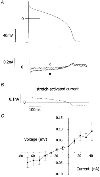
A, upper panel, free action potentials were recorded then used as the waveform to voltage clamp each myocyte. Lower panel, compensation currents (which have a reverse polarity to ionic currents) recorded under action potential clamp before (○) and after (•) a stretch that increased sarcomere length from 1.77 to 2.00 μm. Following decay of large capacitive currents, compensation currents at short sarcomere length are very small. B, stretch-induced current. C, mean current-voltage relationship for the stretch-activated current from 10 cells, having a current of greater than 5 pA at +30 mV. Stretch increased sarcomere length from 1.82 ± 0.01 to 1.98 ± 0.01 μm. The current-voltage relationship was achieved by plotting current (B) against clamp voltage (A, upper panel).