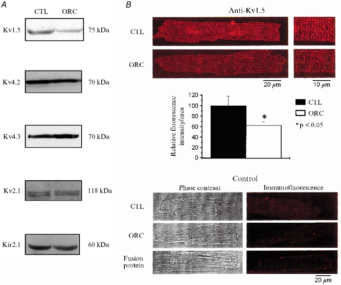
Figure 7. Western blot and immunofluorescence detection of K+ channel expression in CTL and ORC male mouse ventricle.

A, comparison of K+ channel protein expression in CTL and ORC ventricles. Western blot analysis of Kv1.5 (1:500), Kv4.2 (1:500), Kv4.3 (1:4000), Kv2.1 (1:300) and Kir2.1 (1:500) in sarcolemmal-enriched proteins (100 μg lane−1) isolated from CTL and ORC mouse ventricles (n = 2 per group; 3 pooled ventricles per n value). Antibodies used were all obtained from Alomone Labs (Jerusalem, Israel), with the exception of Kv1.5, which was purchased from Upstate Biotechnology (Lake Placid, NY, USA). Equal protein loading was confirmed by Ponceau S-stained membranes. Furthermore, we used Kir2.1 as an internal control on the same Western blot gel as Kv1.5 and found no difference in the density of this protein (data not shown). B, immunofluorescence labelling of Kv1.5 in CTL and ORC male mouse ventricular myocytes. Upper panels (left), isolated cells were stained by exposure to the primary antibody and then to TRITC-conjugated donkey anti-rabbit secondary antibody (Jackson ImmunoResearch Laboratories Inc., Baltimore, PA, USA). The red fluorescence staining indicates the presence of Kv1.5 in CTL and ORC myocytes. Right, cells seen on the left at higher magnification. Middle panel, bar graph showing the relative fluorescence intensity of Kv1.5 in CTL and ORC myocytes (2 mice per group; 10 cells studied per mouse). Individual values of Kv1.5 fluorescence intensity corresponded to whole-cell fluorescence intensity. These values were obtained with the laser scanning microscopy software using an indicator that recorded fluorescence intensity at every pixel of the cell image. These measures were then normalized to cell surface area to account for cell size. Lower panels, phase-contrast images (left) and immunofluorescence detection (right) of the same CTL and ORC cells. These negative controls show that no staining was apparent when the primary antibody was omitted in CTL and ORC cells. The experiment using a fusion protein specific for the sequence of the antibody shows the specificity of the staining for Kv1.5.
