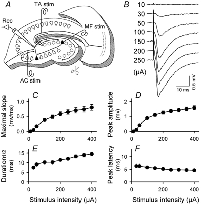
Figure 1. Recording of TA–CA3 fEPSPs.

A, recording set up. A recording electrode was positioned in CA3 stratum lacunosum-moleculare of a slice incised along the sulcus hippocampi and across the edge of the dentate molecular layer (scissors). Three stimulating electrodes were placed in the proximate of the hippocampal fissure, the stratum granulosum and CA3 stratum radiatum to stimulate TA, MF and AC, respectively. B, representative traces of fEPSPs evoked by applying gradually increased intensity of the TA stimulus, ranging from 10 to 250 µA. C-F, input-output relationships of the slope of fEPSPs (C), the amplitude of fEPSPs (D), the width of fEPSPs at half-height (E) and the latency from TA stimulation to reaching the fEPSP peak (F). Almost no shift in the peak latency (only a 21.9 ± 18.7 % decrease) was detected by increasing stimulus intensity, confirming that the fEPSPs reflect TA–CA3 monosynaptic responses. All data represent means ± s.e.m. of 24 slices; where no error bars are apparent they have been obscured by the symbol.
