
Figure 3. Responses of cerebral blood flow (CBF) and cerebral PO2 to 1 h periods of hypoxia and recovery in seven near-term fetal sheep.
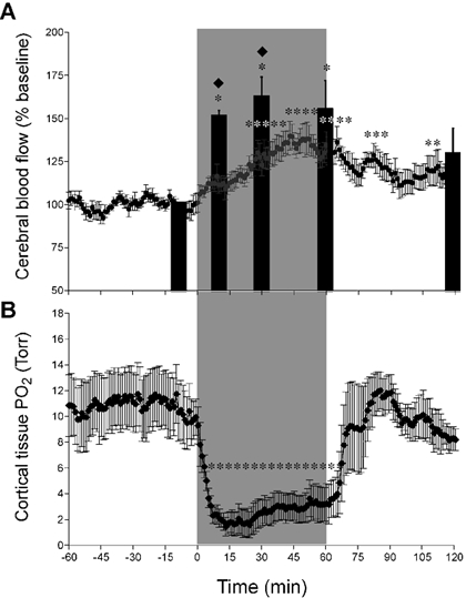
A, results shown as a percentage of baseline; * significantly different from baseline, P < 0.05. Microsphere determinations were made at −10, 10, 30, 60 and 120 min after onset of hypoxia, and results are shown as filled columns. Running 1 min averages of laser Doppler results are shown throughout the experiment. Results of the two methods differed measurably after 10 and 30 min (♦P < 0.05), but were indistinguishable after 60 and 120 min. B, O2 tension in cortical brain tissue in response to a 1 h period of moderate hypoxia followed by a 1 h period of recovery (n = 4). * Significant difference from baseline (P < 0.05). Note the markedly low levels of tPO2 soon after the onset of hypoxia and the increase thereafter in association with the progressive increase of CBF.