
Figure 1. Effects of fasudil and hydroxyfasudil on the prostaglandin F2α (PGF2α)- or 12-deoxyphorbol 13-isobutyrate (DPB)-induced contraction and 20 kDa myosin light chain (MLC20) phosphorylation in rabbit aortae.
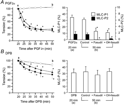
Fasudil (▪) or hydroxyfasudil (OH-fasudil, (Δ) at 10 µm was added 20 min after the application of 10 µm PGF2α (A) or 1 µm DPB (B). Left panels, a change in tension after the addition of fasudil or hydroxyfasudil (○, agonist alone). Tension is expressed as a percentage of contraction just before the addition of fasudil or hydroxyfasudil. In the right panels, MLC20 phosphorylation (MLC-P) was observed 20 min after the addition of PGF2α or DPB (just before the addition of fasudil or hydroxyfasudil (a) or 50 min after the addition of PGF2α or DPB (b), and is expressed as a percentage of phosphorylated MLC20 of total MLC20. MLC20 phosphorylation in the resting state was 8–9 % in each case (data not shown). Since PGF2α caused diphosphorylation of MLC20, monophosphorylation (MLC-P1, open columns) and diphosphorylation (MLC-P2, black columns) are shown (A, right panel). Data are presented as the mean ± s.e.m. (n = 5–6). * Significantly different from the time-matched control (P < 0.05).