
Figure 8. PGF2α-induced phosphorylation of the MBS of phosphatase, and its inhibition by fasudil.
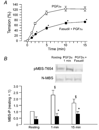
A, time-course of the PGF2α (10 µm)-induced contraction in the absence (○) or presence (•) of fasudil (10 µm). When present, fasudil was applied 15 min before addition of PGF2α. B, (photographs) example of Western blots with the pMBS-T654 antibody or the N-MBS antibody. Samples were taken before the addition of PGF2α (resting) and 1 min after PGF2α. Bar graph, quantified phosphorylation of MBS (MBS-P) in the resting state, 1 or 15 min after the addition of PGF2α in the absence (open columns) or presence (filled columns) of fasudil. The ratio of the density of a band with the pMBS-T654 antibody to that with the N-MBS antibody in the resting state is referred as 1. Data are presented as the means ± s.e.m. of four experiments. * Significantly different from the resting level (P < 0.05); § significantly different from PGF2α alone (P < 0.05).