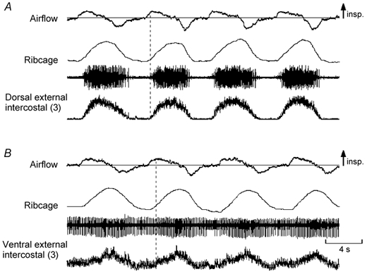
Figure 2. Records from the dorsal and ventral portions of the external intercostal muscle in the third interspace in a subject with inspiratory activity in the ventral portion during quiet breathing.

Same conventions as in Fig. 1. The two portions of the muscle in this subject are active during resting inspiration. However, whereas inspiratory activity in the dorsal portion (A) starts shortly after the onset of inspiratory airflow, inspiratory activity in the ventral portion (B) starts later. In addition, such inspiratory activity is superimposed on tonic activity persisting throughout expiration. Note also that this recording (B) contains far-field contamination during the expiratory pause; this expiratory activity arises from different single motor units, presumably in the internal intercostal muscle, and is associated with a decrease in the end-expiratory position of the ribcage.
