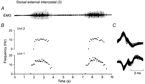
Figure 4. Measurement of the firing rates of single motor units.

A shows a representative record of inspiratory EMG activity obtained in the dorsal portion of the external intercostal muscle in the third interspace for two consecutive breaths. Two single motor units (denoted Units 1 and 2) were clearly identified in this record, and the instantaneous discharge frequencies of these two units are shown in B. All the action potentials from the two units are superimposed in C. Note that all the spikes in C are identical, thus indicating that they originate in a single motor unit. Note also in B that the discharge frequency of both units increases rapidly in the first part of inspiration and plateaus in the last two-thirds of inspiration; the discharge frequency of Unit 1 then declines gradually in the first part of expiration, whereas the discharge frequency of Unit 2 stops rather abruptly after the end of inspiration.
