
Figure 1. Expression of PSD-95 in pyramidal neurons of the prefrontal cortex increases the AMPAR/NMDAR ratio.
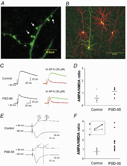
A, confocal image taken after 2 days in vitro illustrating the preferential targeting of PSD-95-GFP fusion protein to dendritic spines (see arrows). B, illustration of the experimental preparation used for these experiments. Prefrontal cortex pyramidal neurons in this slice were transfected with PSD-95/EGFP (green) and (in some cases) with pcDNA3.1/DsRed (red) to serve as within-slice transfected controls. C, current traces recorded at +40 mV from neurons transfected with pcDNA3.1/EGFP (control) or PSD95/EGFP, before (black trace) and after (red trace) administration of 30 µm (d)-AP-5. The green traces depict the NMDAR (AP-5-sensitive) component of the evoked current obtained by subtraction. D, computed AMPAR/NMDAR ratios obtained from PSD-95-transfected neurons (n = 6) and controls (5 cells transfected with pcDNA3.1/EGFP and 3 untransfected neurons; P < 0.02). E, current traces recorded at −60 and +40 mV from a control (pcDNA3.1/EGFP) and from a PSD-95-transfected neuron. By superimposing the traces, the AMPAR (1) and NMDAR (2) components of the traces at +40 mV were estimated. F, computed AMPAR/NMDAR ratios obtained using this method for PSD-95-transfected (n = 14) and control neurons (6 untransfected cells and 7 cells transfected with empty plasmid (i.e. pcDNA3.1/EGFP; sister slice comparisons, n = 5; or pcDNA3.1/DsRed; same slice comparisons, n = 2). A subset of these experiments involving pairwise sequential recordings from neighbouring neurons is re-plotted in the inset. PSD-95 transfection increased the AMPAR/NMDAR ratio (P < 0.01 for pooled data and P < 0.03 for pairwise recordings).