
Figure 1. Dependence of inwardly rectifying currents on extracellular K+ concentration.
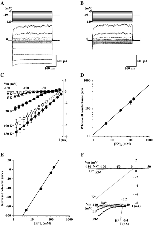
A and B, representative tracings of whole-cell currents obtained from a single bovine parotid acinar (BPA) cells in the presence (A) and the absence (B) of 5 mm K+ (solution B). Hyperpolarizing and depolarizing pulses 400 ms in duration were applied from a holding potential of −49 mV to potentials between −129 and −9 mV in 10 mV intervals. The pipette was filled with a potassium glutamate-rich solution (solution A). C, steady state current-voltage (I-V) relationships of whole-cell currents recorded from BPA cells bathed in different extracellular K+ concentrations. Each point represents the mean ± s.e.m. of 13 experiments. D, log-log plot of the conductance of the inward current as a function of the extracellular K+ concentration; the line is the line of best fit and the data are from C. E, semi-logarithmic plot of the reversal potential of the inward current as a function of extracellular K+ concentration. Each point represents the mean ± s.e.m., but when this was so small as to lie within the symbols, it has been omitted; the line is the line of best fit and the data are from C. F, representative instantaneous I-V relations of whole-cell currents obtained from single BPA cell with Na+, Li+ or Rb+ substituted for K+ (154.3 mm) in the bathing solution (solution C). Ramp command voltages were applied from −125 to +35 mV at a rate of 200 mV s−1. Inset, expanded scale trace for the same experiments.