
Figure 7. Immunofluorescence labelling for Kir2.1 in bovine parotid gland.
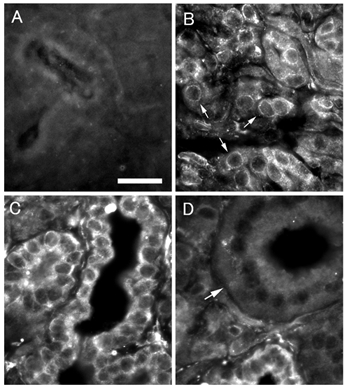
A, control: no labelling is observed. Bar indicates 20 µm. B, acini: peripheries of acinar cells (arrows) are labelled. C, interstitial segment: labelling is observed clearly in entire periphery of interstitial segment cells. D, striated duct: striated duct (arrow) where no labelling is observed.