
Figure 1. Representative photomicrographs of the cerebral neocortex of the fetal baboon.
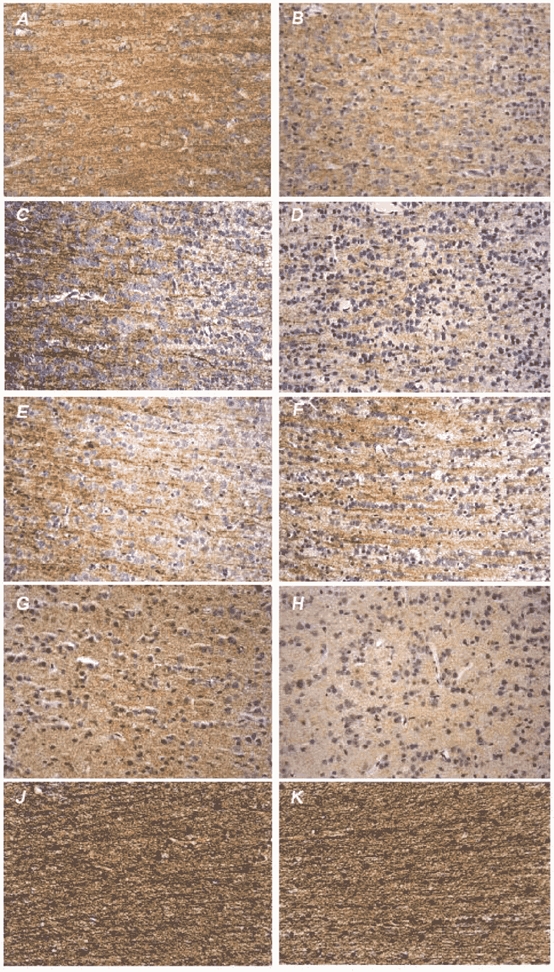
Left: fetus of vehicle-treated pregnant baboon; right: fetus of betamethasone-treated pregnant baboon. Immunohistochemical staining (brown precipitate, hematoxylin counterstaining to visualise cell nuclei) of microtubule-associated proteins MAP1B (A and B), MAP2abc (C and D), MAP2ab (E and F) and presynaptic protein synaptophysin (G and H). J and K, histochemical staining (black precipitate) of argyrophil neurofibrils. All photomicrographs were taken at × 320 magnification. Note the loss of MAP1B, MAP2abc and synaptophysin IR after antenatal betamethasone exposure in the absence of neuronal necrosis. The amount of the high molecular weight isoform MAP2ab remained unchanged. Histochemical staining of neurofibrils of the cortical white matter did not demonstrate effects of betamethasone.