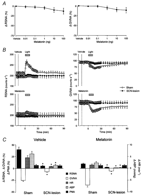
Figure 9. Effects of bilateral SCN lesion on light-induced autonomic and cardiorespiratory responses.

A, group data showing the effects of consecutive i.c.v. injections of melatonin (0.01–100 ng) on RSNA (right, n = 7) and GVNA (left, n = 7) in SCN-lesioned mice. Melatonin (i.c.v.)-induced dose-dependent suppressions of RSNA and GVNA were no longer observed in mice with bilateral SCN lesion (compare these results with those in Fig. 3A and B). B, group data showing time course of the effects of melatonin (0.1 ng) and vehicle i.c.v. injections on light (2.1 × 10¹⁴ photons cm−2 s−1, 10 min)-induced change of RSNA (left panel) and GVNA (right panel) in SCN-lesioned (filled circles, n = 14) and sham-operated (open circles, n = 14) mice. The light-induced RSNA and GVNA responsiveness following i.c.v. injection of melatonin or vehicle observed in sham-operated animals disappeared in SCN-lesioned animals. C, group data showing the effects of SCN lesion on light-induced peak responses of RSNA, GVNA, HR, ABP and PNA in animals pretreated with i.c.v. vehicle (left panel, n = 14) and melatonin (right panel, n = 14). SCN lesion significantly reduced the light-induced increases in RSNA, HR and ABP and decrease in GVNA. *P < 0.05vs. sham-operation.
