
Figure 1. Rapid perfusion of agonist solutions to excised membrane patches.
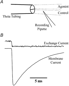
A, schematic diagram of a routine experiment showing the orientation of the theta tubing and recording pipette containing an outside-out membrane patch. To achieve rapid solution exchange of excised patches, both control and agonist solutions were fed continuously by gravity and the tip of the recording pipette was placed close to the control-agonist solution interface. The solution was rapidly exchanged by displacement of the theta tubing using a piezoelectric stack (Physik Instrumente). B, typical exchange and membrane currents recorded from the same patch recording (patch no. 010712p1). The solution exchange rate was determined routinely at the end of each experiment by measuring the current between the solution containing 10 mm l-glutamate and the control solution, in which the total Na+ content was reduced by 5 %.