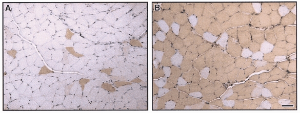
Figure 3. Immunohistochemical analysis of PV expression.

Immunohistochemical staining for PV in soleus (SOL) muscle from wild-type mice and transgenic mice overexpressing the PV-HA transgene. PV was expressed in only a subpopulation of fibres in SOL from wild-type mice (A) and in almost all fibres from TG mice (B). In SOL from TG mice, the positively staining fibres represent both endogenous PV and PV-HA transgene, resulting in ∼6-fold greater number of PV-expressing fibres in TG SOL compared to WT. Bar, 25 μm.
