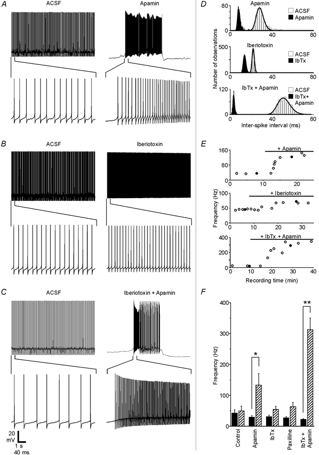
Figure 4. SK and BK channels regulate Purkinje cell firing patterns.

A-C, recordings from three different Purkinje neurons show how spontaneous firing was affected by 200 nm apamin, 100 nm iberiotoxin or the combination of the two blockers. The same scale bar applies to each set of traces. D, ISI distributions and fits are shown for the traces in A-C. E, spontaneous firing frequency plotted as a function of recording time for each experiment. •, the traces shown in A-C. Black horizontal bars show when toxins were present in the bath solution. Response delays partly reflect the time needed for residual control solution to empty out of the perfusion tubing and inline solution heater. F, the effects of specific KCa channel blockers on the firing frequency are summarized. Black bars show the baseline frequency for each group (means ±s.e.m.), while hatched bars show the frequency in the presence of the blocker. The firing frequency increased from 29 ± 5 to 133 ± 37 Hz with the application of apamin (n = 7 cells), from 29 ± 5 to 50 ± 12 Hz with iberiotoxin (n = 6), from 27 ± 4 to 63 ± 14 Hz with 1 μm paxilline (n = 5) and from 22 ± 3 to 313 ± 37 Hz when both iberiotoxin and apamin were applied together (n = 5). For the control group, the firing rate increased from 41 ± 16 initially to 47 ± 19 Hz after 10 min of control recording time (n = 6). Apamin had a significant effect compared to the control (P < 0.05), and the simultaneous application of iberiotoxin and apamin differed significantly from both the control group (P < 0.0001) and from the apamin group (P < 0.01). When applied alone, neither iberiotoxin nor paxilline had a statistically significant effect compared to the control group (P > 0.5).
