
Figure 1. Expiratory carbon dioxide concentrations, R–R intervals, and a horizontal section through a sliding fast Fourier transformation of R–R intervals during ramped frequency breathing.
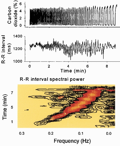
Respiratory sinus arrhythmia (middle and bottom panels, black to orange areas) was small at the extremes of breathing frequency. Low frequency spectral power (bottom panel, extreme right) was present throughout the period of ramped breathing. These data indicate that some R–R interval fluctuations closely track breathing.