
Figure 2. 13C spectra from rested and contracted gastrocnemius and soleus muscles.
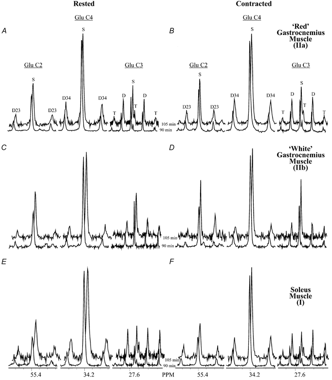
Glutamate carbon 2 (C2), C4 and C3 resonances are shown from 13C spectra of representative extracts of rested and contracted muscles infused with [2,4,6,8-13C4]octanoate for 90 or 105 min. A and B, predominantly red rested (A) and contracted (B) gastrocnemius muscle (IIa); C and D, rested (C) and contracted (D) white gastrocnemius muscle (IIb); E and F, rested (E) and contracted (F) soleus muscle (I). Visible multiplets are as follows: the C2 singlet (S) and the doublet resulting from carbon-carbon coupling of carbons 2 and 3 (D23) in the glutamate C2 region, the C4S and doublet 34 (D34) in the glutamate C4 area, and the S, D and triplet (T) in the glutamate C3 region.