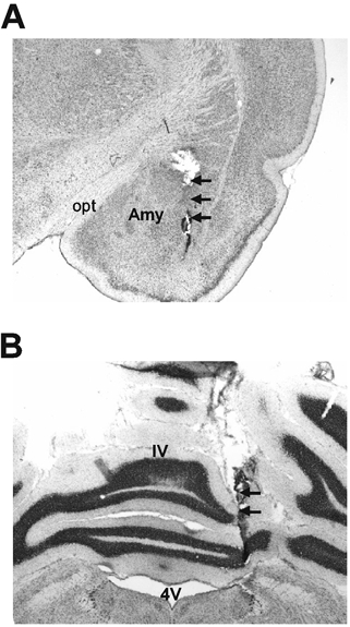
Figure 5. Cresyl-violet-stained coronal brain section showing location of the microdialysis probes.

A, the amygdala. B, the cerebellar cortex. The locations of the microdialysis probes are indicated by arrows. Opt: optic tract; Amy: central nucleus of amygdala; 4V: 4th ventricle; IV: preculminate fissure 4.
