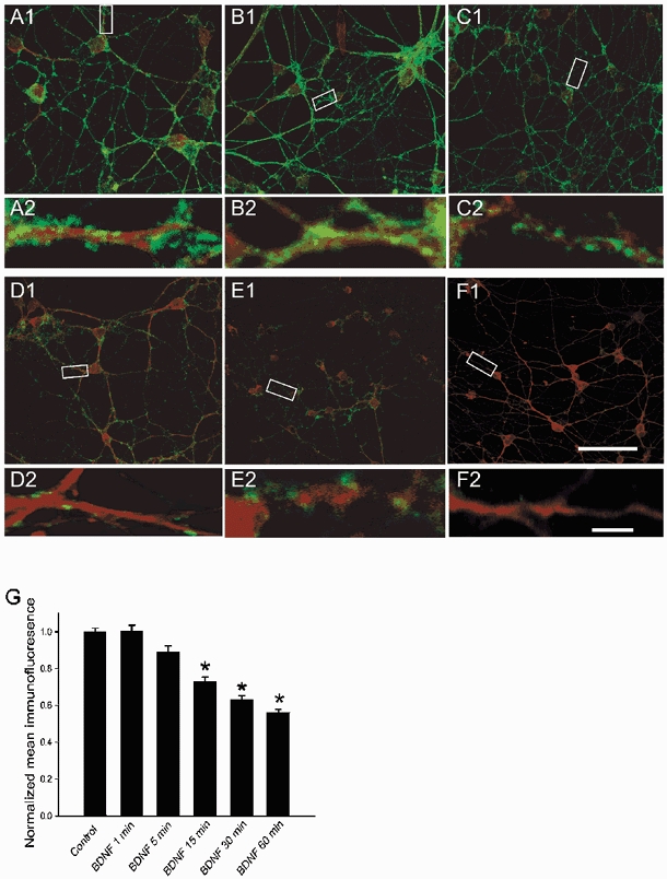
Figure 5. Exposure to BDNF reduces GABAA receptor β2/3 subunit immunoreactivity in a time-dependent manner.

Confocal images of cultured cerebellar granule cells before (A1) and following a 1 (B1), 5 (C1), 15 (D1), 30 (E1) and 60 (F1) min exposure to BDNF (100 ng ml−1). The cultures were fixed and double-labelled with antibodies against GABAA receptor β2/3 subunits (green fluorescence) and TrkB receptors (red fluorescence). A2, B2, C2, D2, E2 and F2, GABAA receptor β2/3 subunit immunoreactive profiles corresponding to the boxed areas in A1-F1 are resolved at higher resolution (× 60 oil) to illustrate decreased immunoreactivity along neurites following a > 5 min exposure to BDNF. Scale bars, 50 μm in A1-F1 and 5 μm in A2-F2. The images illustrated above were digitized; relative median density level was determined for each treatment and normalized to control. G, data are presented as means ±s.e.m. * Significant difference between control and the corresponding times of exposure to BDNF (100 ng ml−1) (Student's t test at P < 0.01).
