
Figure 4. 135 mm Cs0+ slows both onset of and recovery from HERG inactivation.
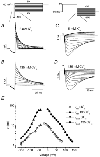
A and B, HERG currents elicited by the voltage protocol shown above the current traces in external solution containing 5 mm K0+ (A) or 135 mm Cs0+ (B) with an internal solution containing 130 mm K+. Starting from a holding potential of +60 mV, a 10 ms repolarizing step to −100 mV allowed HERG channels to recover from inactivation, and steps to a range of test potentials allowed the inactivation time course to be observed. Current traces represent responses in the time interval demarcated by the slanted lines in the voltage protocol. C and D, the time and voltage dependence of recovery from inactivation was measured by the voltage protocol shown above the current traces in external solution containing 5 mm K0+ (C) or 135 mm Cs0+ (D) and with 130 mm K1+. HERG was activated by a 400 ms pulse to 60 mV, followed by a test pulse to different potentials to record HERG tail currents. The time constant of recovery (τrec) from inactivation was measured as the single exponential fit to the rising phase (> −80 mV) or as the fast time constant of a double exponential fit (≤−80 mV) to HERG tail current (Sanguinetti et al. 1995; Spector et al. 1996). E, voltage dependence of the τrec and the inactivation time constant (τinact) in external solution containing 5 mm K0+ (▵, O) or 135 mm Cs0+ (▴, •). Each point is the mean ±s.e.m. for eight to nine cells.