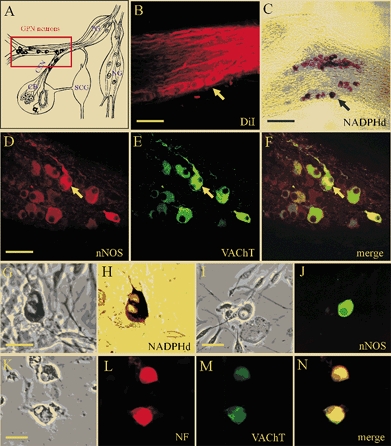
Figure 1. Localisation and properties of neurones of the rat glossopharyngeal nerve (GPN) in situ and in culture.

A, schematic drawing of the carotid bifurcation showing localisation of GPN neurones (red rectangle) embedded in the glossopharyngeal and carotid sinus (CSN) nerves (modified from Wang et al. 1993). Petrosal ganglion (PG), nodose ganglion (NG), superior cervical ganglion (SCG) and carotid body (CB) are indicated in the diagram. B, the GPN neurones (yellow arrow) are retrogradely labelled following injection of the lipophilic dye DiI (1,1′-dioctadecyl-3,3,3′,3′-tetramethylindocarbocyanine perchlorate) into the CB; whole-mount of glossopharyngeal nerve visualised with confocal microscopy. C, GPN neurones (black arrow) are visualised in situ in a whole-mount of glossopharyngeal nerve after staining for NADPH-diaphorase (NADPHd) activity. D-F, same microscopic field of GPN neurones in situ showing positive immunostaining for nNOS (D; Cy3) and the cholinergic marker VAChT (E; fluorescein isothiocyanate (FITC)); note co-localisation of the two markers (e.g. yellow arrow) in the same neurones (F). G and H, corresponding phase and brightfield images of positive NADPHd activity in two adjacent neurones after 2 days in culture. I and J, corresponding phase and nNOS immunofluorescence (FITC) in a cultured GPN neurone, 2 days after isolation. K-N, corresponding phase and fluorescence micrographs showing a pair of cultured GPN neurones that were immunopositive for both neurofilament (NF, L; Alexa 594) and VAChT (M; FITC); overlap of the two markers is shown in N. Scale bars represent 50 μm in B and D; 150 μm in C and 30 μm in G, I and K.
