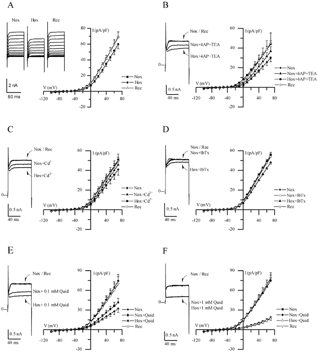
Figure 2. Electrophysiological responses of cultured GPN neurones to hypoxia.

A, traces show a representative example of the reversible reduction of macroscopic outward current recorded in a GPN neurone after exposure to hypoxia in physiological extracellular K+ (5 mm) solution, during various step potentials from −100 to +70 mV. Current-voltage (I-V) plots of the current density (mean ±s.e.m.; n = 11) during normoxia (Nox), hypoxia (Hox) and recovery (Rec) are shown on the right; hypoxia caused a significant reduction in current density (P < 0.05) at potentials > 30 mV. B, traces show a representative example of the response to hypoxia in physiological solution containing 4-AP (2 mm) and TEA (5 mm), during a step to +40 mV; I-V plots show mean ±s.e.m. (n = 6) current density during normoxia and hypoxia in the presence of 4-AP and TEA. Note that hypoxic inhibition of the macroscopic outward currents persisted in physiological solution containing these classical voltage-dependent K+ channel blockers. Similar results were obtained in the presence of 200 μm Cd2+ (n = 6), 100 nm IbTx (n = 5) and 0.1 mm quinidine (Quid: n = 5) as shown in C, D and E, respectively. However, as shown in F, higher concentrations of quinidine (1 mm) caused further suppression of the outward current and, moreover, abolished the response to hypoxia (n = 7).
