
Figure 2. Responses mediated by GluR5-containing KARs are modulated in a PKC-dependent manner.
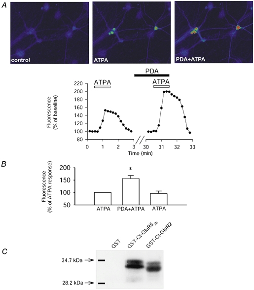
A, single example of a field of neurones (top) and the analysed change in fluorescence (bottom) showing that application of ATPA (2 μm), to selectively stimulate GluR5-containing KARs, results in an increase in fluorescence of the calcium indicator fluo-3 AM. The co-application of phorbol esters (PDA; 1 μm), to stimulate PKC, enhanced the GluR5-mediated calcium rise. B, pooled data showing that ATPA-induced calcium signalling is significantly increased by stimulation of PKC in a reversible manner (n = 18 cells, 3 dishes; P < 0.01). Following washout of PDA, the response to ATPA returns to baseline levels (96 ± 10 %). C, GluR52b is phosphorylated by PKC. A single representative example showing that GST (n = 10) is not phosphorylated by PKC. However, GST-Ct-GluR52b (n = 10) and GST-Ct-GluR2 (n = 2) are both phosphorylated by purified PKC.