
Figure 2. Sagittal-plane scans of the patella tendon (PT) at rest, during isometric contraction at 50 % of maximal and at maximal tendon force.
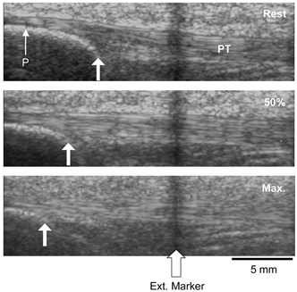
Arrows indicate displacement of the apex of the patella (P) during contraction with respect to an echo-absorptive external marker fixed on the skin.