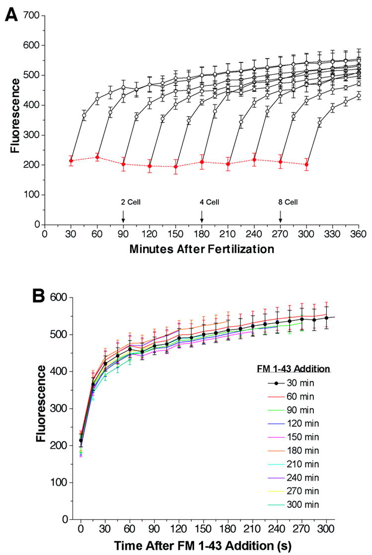
Figure 1.

The instantaneous and cumulative FM1-43 fluorescence observed during early development. Panel A: A 5% suspension of sea urchin eggs (Lytechinus pictus) was fertilized with sperm (0.5 μl/ml) in a single pool and aliquots of the fertilized eggs were added to wells of a low fluorescence microtiter dish in triplicate for each time point and kept at 16°C. At 30-minute intervals after fertilization FM1-43 was added to the wells at a final concentration of 4 μM to measure the embryo surface area and membrane flux. Fluorescence (Ex 480/Em 580) was determined immediately (‘Instantaneous’ in RED) and at 15 min intervals (‘Cumulative’ in BLACK). Between readings, eggs were kept at 16°C. Indications of cell-stage number are shown when ~90% of the eggs were at that stage. All points are mean ± SEM of three experiments. Panel B: Traces in panel A are overlaid by plotting them as a function of time after FM1-43 addition.
