
Figure 5.
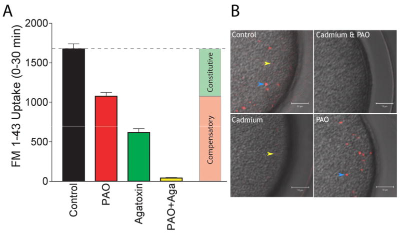
Two forms of endocytosis account for virtually all of the uptake observed during early development. Panel A: A suspension of sea urchin eggs (Strongylocentrotus purpuratus) were treated with either 10 μM PAO (red bar), 100 nM agatoxin-TK (green bar), or a combination of both inhibitors (yellow bar) and FM1-43 uptake during a 30 minute period following fertilization was measured and compared with control eggs (black bar). PAO was added 2 minutes after the addition of sperm, along with 4 μM FM1-43. Agatoxin was added at the same time as sperm and FM1-43. When both PAO and agatoxin were used the agatoxin and sperm first added, followed 2 minutes later by the addition of PAO and FM1-43. Control eggs were treated with sperm and FM1-43 at the same time. 30 minute after the addition of FM1-43 eggs were washed in ASW containing 1 mg/ml trypan blue (to quench any tightly bound extracellular FM1-43 fluorescence), resuspended in ASW, and aliquots transferred to a microtiter plate for fluorescence determination. Each point represents data in triplicate from three separate animals and is the mean ± SEM, n=3. Panel B: Two-photon microscopy was used to image the uptake of a fluorescent fluid phase marker (100 μM tetramethylrhodamine-dextran mol. wt. 3000, TMR-dex) in fertilized eggs (Strongylocentrotus purpuratus), or eggs treated with cadmium (500 μM), PAO (10 μM), or a mixture of cadmium and PAO. Eggs were fertilized with sperm, and at 2 minutes after fertilization inhibitors were added along with the TMR-dex. Eggs were washed with ASW 30 minutes later (to remove extracellular TMR-dex) and imaged. Images are an overlay of the two-photon TMR-dex fluorescence image and the laser-scanning DIC image. Yellow arrow head indicates the small endosomes formed by the PAO sensitive form of endocytosis and the blue arrow heads indicates the large endosomes formed by the cadmium sensitive form of endocytosis. Size bars are 10 μm.