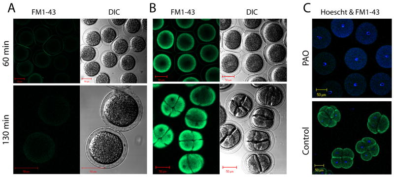
Figure 7.
PAO inhibits cell division. Fertilized eggs (Strongylocentrotus purpuratus) were either incubated for 10 minutes with 10 μM PAO (panel A), or mock treated (panel B), and then incubated in ASW containing FM1-43 (4 μM) for 30 minutes. Eggs were next washed with ASW to remove extracellular FM1-43 and imaged with two-photon microscopy at 60 and 130 minutes post fertilization to image both cell division (by DIC) and FM1-43 uptake (green fluorescence). Next, both control embryos and those treated with PAO were stained with Hoechst stain (20 μg/ml) for 15 minutes, washed with ASW and cell nuclei were visualized (see blue fluorescence in panel C) with two-photon microscopy. Size bars are 50 μm.

