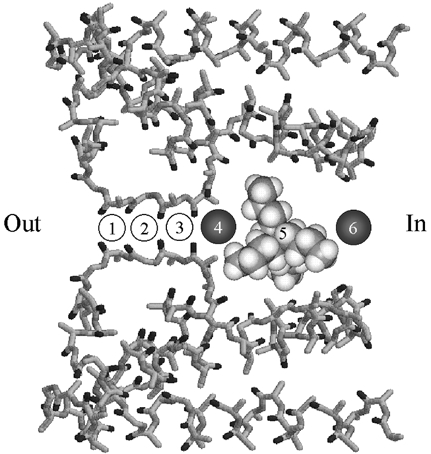
Figure 9. Relative location of ion binding sites in the inner end of the pore in Shaker K+ channels.

Possible sites of ion binding in Shaker K channels superimposed on the pore structure of the MthK channel (PDB entry 1LNQ) (Jiang et al. 2002). Sites labelled 1 to 6 are described in the text. Site 5 is shown occupied by a space-filling model of TBSb. This space-filling model is not intended to represent a particular structure for TBSb but only serves as an indication of its relative size. Circles occupying sites 4 and 6 represent K+ ions and are approximately to scale with the space-filling model. ‘Out’ and ‘In’ represent the orientation of the diagram with respect to the external and cytoplasmic ends of the pore, respectively.
