
Figure 9. Multiple single-channel conductances of somatic AMPARs in Purkinje cell patches.
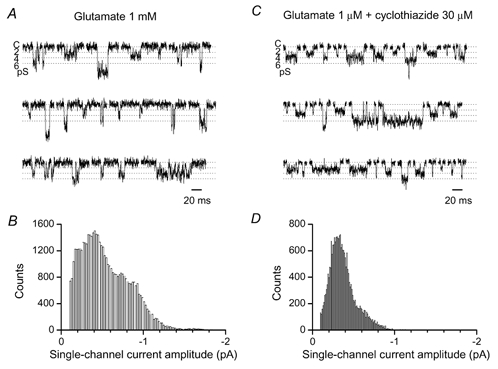
A and C, examples of single-channel openings in the continued presence of agonist, recorded from outside-out patches of P3 Purkinje cells. Vh, −100 mV. External Ca2+, 5 mm. Baseline (closed) level is indicated as C. B and D, all-point amplitude histograms of single AMPA channel currents. The baseline noise has been subtracted from histograms after fitting Gaussian distributions to the noise components.