
Figure 2. Renal morphology in adult control and prenatally dexamethasone-treated animals.
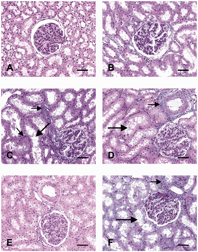
Representative photomicrographs of the renal cortex from two animals from the control group (CON; A and B) and four animals prenatally treated with dexamethasone (DEX; C–F); Tissues shown in A and E were stained with haematoxylin and eosin and those in B, C, D and F were stained with Masson trichrome. In three out of four DEX animals (C, D and F) the proximal tubules (large arrows) were markedly dilated and enlarged. There was no noticeable collagen accumulation in the glomeruli of any animal, but excess collagen can be seen in the tubular interstitium and the periadventitia of cortical vessels (small arrows). Bar represents 50 μm.