
Figure 3. Kidney dry weight, total collagen content and collagen types (I and III) in adult control and prenatally dexamethasone-treated animals.
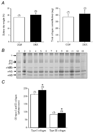
A, the total kidney dry weight, expressed as a percentage of the wet weight of tissue and total kidney collagen content (converted from the hydroxyproline values) from the control group (CON; □ n = 7) and dexamethasone-treated group of animals (DEX; ▪; n = 5). B, the pepsin-digested collagen (which represents the maturely cross-linked collagen) was analysed by SDS-PAGE, using delayed reduction of the disulphide bonds with 10% β-mercaptoethanol. The samples consist of a type I and type III collagen standard (from human dermis) (lane 1) and the pepsin digests of renal tissues from the CON (lanes 2–8) and the DEX group of animals (lanes 9–13). Type I collagen is normally composed of two α1(I) chains and one α2(I) chain, while type III collagen is composed of three α1(III) chains. β11 and β12 represent dimers of two μ1(I) chains and an α1(I)/α2(I) chain, respectively, while γ represents collagen trimers. C, the optical density (OD) of the type I and type III collagen chains (from the individual samples of kidneys taken from the CON (□;) and DEX (▪;) groups of animals was determined using a Bio-Rad GS-710 Calibrated Imaging Densitometer. The results from the individual samples were then grouped. *P < 0.05, when compared to OD values from the CON group.