
Figure 4. Effects of EGF and PDGF peptides on whole-cell VSOR Cl− currents in control (left panel) and BPV-transfected C127 cells (right panel).
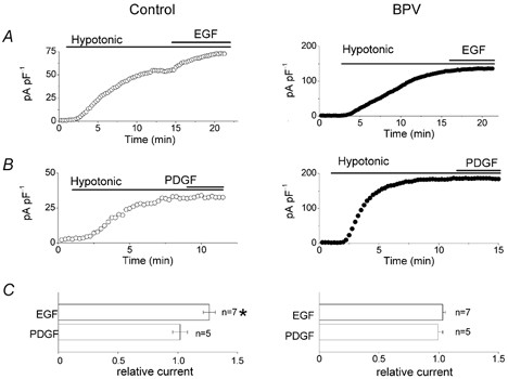
A and B, effects of EGF (50 ng ml−1) and PDGF (20 ng ml−1) on whole-cell VSOR current densities recorded at +40 mV in swollen control and BPV cells. C, summary of these effects. The current densities were normalised to the values recorded before application of EGF or PDGF. * Significantly different from the control values recorded before application of EGF or PDGF.