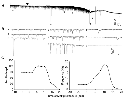
Figure 1. MeHg-induced changes in amplitude and frequency of sIPSCs in a cerebellar Purkinje cell in slice.

A, continuous recording of the time course of effects of bath-applied MeHg (100 μM) on spontaneous inhibitory currents (sIPSCs) recorded from a representative Purkinje cell in a slice at a holding potential of −60 mV. Note, the arrowhead indicates the starting point of MeHg exposure. Prior to that time, the cell had been held continuously for approximately 15 min to ensure that the recording was stable (only ≈5 min trace is shown here). All recordings were made in the presence of CNQX (10 μM) and APV (50 μM) in the external solution to block glutamatergic synaptic function. The lower case letters (a-i) indicate specific changes in spontaneous events before and during MeHg exposure. B, the same sampling points as in A are shown on an expanded time scale: a and b, control; c, g and h, MeHg-induced giant slow inward currents; d and e, MeHg-induced initial peak increase in sIPSC amplitude and frequency; f, spontaneous repetitive firing of Purkinje cell; i, complete cessation of whole-cell currents. C, time courses of effects of MeHg on Purkinje cell sIPSC amplitude (left) and frequency (right). MeHg was applied at time = 0 min. Data were sampled over 2 min periods and represent the average for that 2 min block.
