
Figure 2. MeHg causes a biphasic effect on frequency and amplitude of sIPSCs of cerebellar Purkinje cells which is concentration and time dependent.
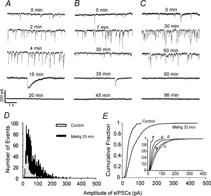
sIPSCs were recorded from Purkinje cells in three sagittal cerebellar slices, following continuous perfusion with MeHg at 100 (A), 20 (B) and 10 μM (C). The holding potential was −60 mV, and all recordings were made in the presence of 10 μM CNQX and 50 μM APV in the external solution. A-C, time courses of effects of MeHg on sIPSCs. Data were collected before (control) and at different time points after exposure to MeHg. Each trace is a representative depiction of 5–11 individual experiments. D, histogram of amplitude distribution of sIPSCs recorded from a Purkinje cell before (control, open bar) and after exposure to 10 μM MeHg for 33 min (filled bar). E, cumulative amplitude distribution of sIPSCs in control (220 events) and at 30 min after exposure to 10 μM MeHg (1252 events). MeHg shifted the cumulative amplitude distribution curve to the right after exposure for 30 min (P < 0.05, Kolmogorov-Smirnov test). Inset, 10 μM MeHg-induced shifting of the cumulative amplitude distribution curves in the same cell at different time points of exposure: a, control, 220 events; b, 15 min, 645 events; c, 30 min, 1252 events; d, 60 min, 747 events; e 90 min, 76 events.