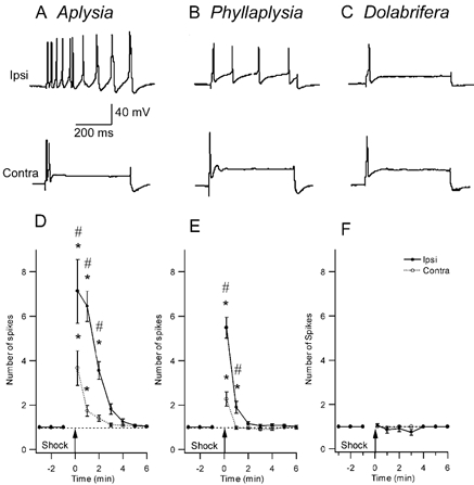
Figure 3. Comparison of the effect of nerve shock on excitability of tail sensory neurons in the three study species.

A-C, representative examples of intracellular recordings from homologous SNs in three species. The sensory neuron was injected with a 500 ms intracellular pulse of depolarizing current, 1 min after electrical stimulation of a peripheral tail nerve (top trace: ipsilateral tail-nerve shock; bottom trace: contralateral tail-nerve shock). Current amplitude was set prior to nerve stimulation to give a single action potential (traces not shown). D-F, SN excitability across time after nerve stimulation shows the change in the number of evoked spikes after nerve shock. D, Aplysia (n = 17) shows a significant increase in excitability both ipsi- and contralateral to the shock, that lasts about 3–5 min. E, in Phyllaplysia (n = 17), nerve shock increased excitability only in the ipsilateral SNs for 1 min after nerve stimulation. F, in Dolabrifera (n = 11), no excitability increase was observed after nerve shock. * Significant difference between pre- and post-test values (P < 0.05); # significant difference between ipsilateral and contralateral sides (P < 0.05).
