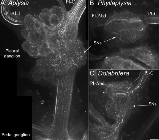
Figure 4. Immunohistochemical labelling of serotonin-immunoreactive fibres in the vicinity of tail sensory neurons.

Confocal photomicrographs of clusters of tail SNs in the pleural ganglion, stained with an anti-5-HT antibody were taken from Aplysia (A), Phyllaplysia (B) and Dolabrifera (C, n = 2). The orientation is the same for all species: pleural ganglion (top), pedal ganglion (bottom), pleural-abdominal nerve (Pl-Abd, left) and pleural-cerebral nerve (Pl-C, right). The three species show a similar pattern of 5-HT innervation: numerous 5-HT fibres are present in the cerebral-pleural nerve, they branch over the cluster of tail SNs (arrows) and/or travel to the abdominal ganglion through the pleural-abdominal nerve. Some 5-HT processes also travel towards the pedal ganglion through the pleural-pedal nerve. No 5-HT-immunoreactive cell bodies were evidenced in the pleural ganglion. Scale bar = 100 μm.
