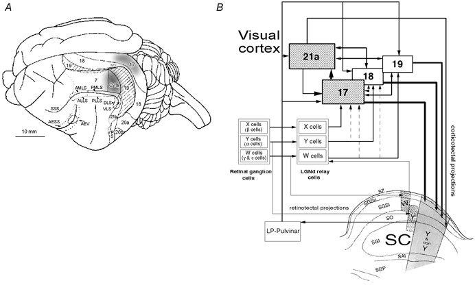
Figure 1. Visual cortical areas and principal connections of superior colliculus of the cat.

A, dorsolateral view of the surface anatomy of the left cerebral hemisphere of the cat with cortical visual areas outlined. In addition to visuotopically organised cytoarchitectonic areas 7, 17, 18, 19, 21a, 21b, 20a and 20b there are several visuotopically organised lateral suprasylvian areas (anteromedial, anterolateral, posteromedial, posterolateral, dorsal and ventral; AMLS, ALLS, PMLS, PLLS, DLS and VLS, respectively), posterior suprasylvian (PS) and anterior ectosylvian visual area (AEV). AESS, anterior ectosylvian sulcus: MS, marginal sulcus; SSS, suprasylvian sulcus. The borders outlined by interrupted lines are buried in sulci (after Palmer et al. 1978; Tusa et al. 1978, 1979; Updyke, 1986). The shaded part of area 17 corresponds visuotopically to area 21a and the part of the superior colliculus we recorded from. Location of cytoarchitectonic area 7 is also indicated. Area 21a, or the shaded part of area 17 were briefly inactivated by cooling them to 10 °C. When one of the areas was cooled the other was warmed to 36 °C. B, simplified neuronal circuitry of retino-geniculo-cortical, retino-collicular, cortico-cortical and cortico-tectal pathways. A schematic diagram of a coronal section through the superior colliculus (SC) is also incorporated in the diagram. The anatomical subdivisions of the SC are: SZ, stratum zonale; SGSu, stratum griseum superficiale upper; SGSl, stratum griseum superficiale lower; SO, stratum opticum; SGI, stratum griseum intermediale; SAI, stratum album intermediale; SGP, stratum griseum profundum. Note that while the dorsal lateral geniculate nucleus (LGNd) and indirectly the visual cortices are innervated by three distinct functional and morphological types of retinal ganglion cells (X, Y, W), the SC is innervated only by Y and W-type retinal ganglion cells. The primary visual cortices (areas 17 and 18) and area 19 all receive direct W-type geniculate input. Direct Y-type geniculate input is restricted to areas 18 and 17 while direct X-type geniculate input is restricted to area 17. Area 21a constitutes part of the form/pattern- rather than motion-processing stream (cf. Burke et al. 1998; Lomber, 2001). The superficial layers of the SC project to laminae C of the LGNd (containing W cells) as well as to the lateral posterior–pulvinar complex (LP-pulvinar) of the dorsal thalamus which in turn projects to cortical areas 17, 18, 19 and 21a. The cortico-tectal projections from primary visual cortices are more numerous and tend to terminate more superficially than those from areas 19 and 21a. Furthermore, some terminals from areas 19 and 21a, unlike these from areas 17 and 18, terminate in the SGI (after Dreher, 1986; Berson, 1988; Harting et al. 1992; Dreher et al. 1996; Burke et al. 1998; Waleszczyk et al. 1999).
