
Figure 9. Measurements of GABA action on the cell membrane potential using cell-attached recordings.
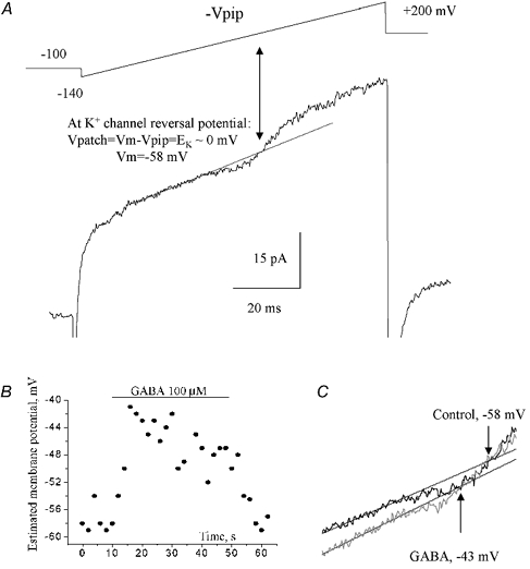
A, a cell-attached record in response to a depolarizing ramp command (shown above the trace) from −140 to +200 mV (-Vpip). The holding potential (-Vpip) was −100 mV with respect to the cell membrane potential. Voltage-gated K+ currents activated by the depolarizing voltage ramp were initially inward, and then reversed to become outward. EK was determined from the intersection of the fit (straight line) to the linear leak and the K+ current. The cell resting membrane potential was measured from the reversal of cell-attached K+ currents. B, plot of the cell membrane potential measured as shown in A against the recording time. GABA was pressure applied for 40 s as indicated by the bar. C, determination of K+ current reversal before and during GABA application (6 s after the start of the application).