
Figure 3. Nav1.8-LI intensity and conduction velocity.
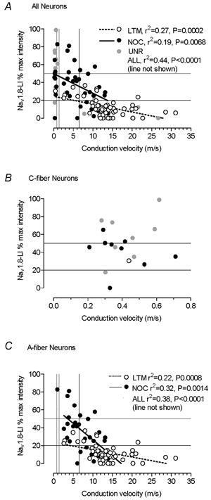
Nav1.8-LI intensity plotted against dorsal root CVs for all DRG neurons examined (A) and for all A-fibre neurons (B). The P and r2 values are given only if the correlation is significant (P < 0.05). Both the borderline between Nav1.8-LI-positive and -negative neurons (20 % maximum intensity) and 50 % maximum intensity are shown by lines on the y-axis. The lines from the x-axes show the C (0.8 m s−1), C/Aδ (1.4 m s−1) and the Aδ/Aα/β (6.5 m s−1) borderlines. NOC, nociceptive neurons; LTM, low threshold mechanoreceptive neurons; UNR, unresponsive neurons.