
Figure 5. Examples of Nav1.8-LI in identified neurons.
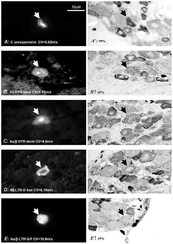
Shown on the left are 5 DRG neurons injected with a fluorescent dye (marked with arrowheads) and on the right are the same neurons stained for Nav1.8-LI. Their sensory properties, dorsal root CV (bottom left) and Nav1.8-LI relative intensity (bottom right) are given. Neurons were considered positive if their Nav1.8-LI was ≥ 20 % and negative if it was < 20 %. HTM, high threshold mechanoreceptive neurons; CV, conduction velocity; LY, Lucifer yellow; CB, Cascade blue.