
Figure 6. Examples of the immunohistochemical staining used.
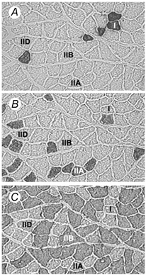
Staining was used to identify: A, type I (anti-MHCI, clone NOQ7.5.4D); B, type IIA (anti-MHCIIA, clone SC-71); C, type IIB (anti-MHCIIB, clone BF-F3) fibres. There were no fibres present which expressed MHC-embryonic. Note that the fibre types marked I, IIA and IIB were identified by their reaction with the corresponding MHC antibody, while IID/X fibres (denoted IID), were identified by the absence of staining.