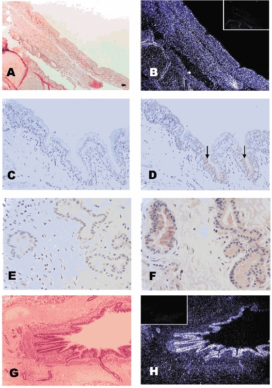
Figure 2. Localization of AQP4,5 in adult lung.

Light (A) and dark (B) field pictures of adult sheep lung, probed with a riboprobe to AQP5. In the adult lung, AQP5 mRNA (white dots) was present in the epithelial cells of the bronchus and in the glands of the submucosa (B). Similarly, the AQP5 protein was located at the apex of the ciliated columnar epithelial cells (D) and in the membranes of the bronchial glands (acinar cells, F) in adult lung tissue; C and E are negative controls for AQP5 immunohistochemistry. Light (G) and darkfield (H) pictures of an adult lung bronchiole demonstrate the distribution of mRNA for AQP4, with a strong signal found in the columnar epithelial cells (H); inserts in B and H are negative controls, using the sense probe. Magnification: A and B (× 100); C and D (× 200); E and F (× 400); G and H (× 40).
