
Figure 1. Experimental apparatus and study protocols.
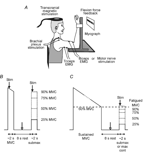
A, experimental apparatus. B, study 1 protocol. Subjects performed pairs of contractions involving a brief maximal voluntary contraction (MVC) followed by a brief submaximal contraction without fatigue. Transcranial magnetic stimulation of the motor cortex (TMS), electrical stimulation of the brachial plexus, or electrical stimulation of the biceps brachii motor nerve was delivered during each contraction (filled arrows). In some contraction pairs, electrical stimulation of the biceps brachii motor nerve was also delivered at rest between contractions (open arrow). C, study 2 protocol. Subjects performed pairs of contractions of the fatigued elbow flexor muscles. Each contraction pair involved a sustained MVC (maintained until maximal force had decreased to 60 % of maximal force without fatigue) followed by a brief maximal or submaximal contraction. Arrows indicate the timing of the stimuli.