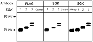
Figure 1. Specificity of the anti-sgk antibody and identification of sgk isoforms expressed in the kidney.

Western blots of proteins extracted from HEK 293 cells transfected with sgk1, sgk2 and sgk3 cDNAs, control (non-transfected cells), and renal tissue examined with anti-FLAG monoclonal and anti-sgk antibodies. The lane labelled kidney contains 5-fold more protein than the lanes loaded with HEK 293 cell lysates. The arrows on the left indicate molecular masses of standards.
