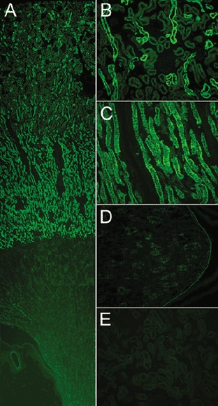
Figure 2. Distribution of sgk1 in kidneys from normal rats examined by immunohistochemistry with anti-sgk antibody.

A, low magnification of a sagittal view of the kidney shows cortex, medulla and papilla. B, higher magnification of cortex; immunoreactivity is detected in distal tubules but not in proximal tubules or glomeruli. C, outer medulla; fluorescent signal localizes to arrays of tubules formed by tall cells, consistent with the thick ascending limb (TAL). There is a marked and sharp decrease in immunoreactivity in the transition from outer to inner medulla. D, papilla. A line was drawn to visualize the limit of the papilla. E, competition of primary antibody with the cognate peptide eliminates fluorescent signal from the renal cortex.
