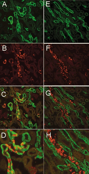
Figure 4. Double-labelling of sgk1 and AE1.

The left and right panels show images from renal cortex and outer medulla, respectively. A, cortex stained with sgk. B, AE1 shows staining of basolateral membranes of intercalated cells in CCT. C, overlay of A and B shows colocalization of sgk1 and AE1 in CNT and CCT. D, larger magnification of a CCT with intercalated cells stained with AE1 and principal cells with sgk1. Outer medulla stained with sgk1 (E), AE1 (F), overlay of E and F (G). H, higher magnification of G shows that sgk1 is not expressed in MCT.
