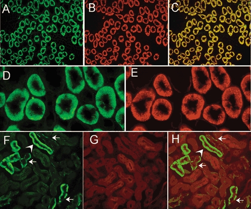
Figure 8. Double-staining of kidney sections for sgk1, the Na+,K+-ATPase and actin.

Staining of a transverse section of the outer medulla for sgk1 (A), the α subunit of the Na+,K+-ATPase (B), and overlay of these two images (C) showing colocalization of the two proteins. Higher magnification of the outer medulla stained for sgk1 (D), and the α subunit of the Na+,K+-ATPase (E) are also shown. Deep infolds of the basolateral membrane are delineated by the two antibodies whereas no fluorescent signal is apparent in the apical membrane. F, section of cortex stained with anti-sgk1. Arrows indicate the basolateral localization of the signal. These tubules correspond to CCT because the staining is restricted to principal cells and absent in intercalated cells. Actin labelled with DNase I conjugated with Texas red (G) distributes over the whole cytoplasm with enhancement of apical microvilli. Overlay shows sgk1 in the basolateral membrane but absent from the apical membrane (H).
