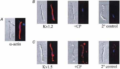
Figure 6. Normarski and corresponding fluorescent images of freshly isolated rat cerebral VSMCs.

A, expression of smooth muscle-specific α-actin. Cells were visualized using Alexa Fluor 594-conjugated antibody (in red). Nuclei were labelled with DAPI (in blue). B, left to right, VSMCs labelled with anti-KV1.2 (Kv1.2), after adsorption of anti-KV1.2 with a competing peptide (+CP) or after incubation with secondary antibody only (2 ° control). C, left to right, VSMCs labelled with anti-KV1.5 (Kv1.5), after adsorption of anti-KV1.5 by a competing peptide (+CP), and after incubation with secondary antibody only (2 ° control). Within each panel, fluorescent images were acquired from VSMCs exposed for equivalent times. Results were verified in 3 (α-actin), 9 (Kv1.2) and 5 (Kv1.5) cells from different preparations.
