
Figure 5. Modulation of α,β-methylene ATP (αβme-ATP)-activated P2X receptor currents by DHEA, ivermectin and low pH solution.
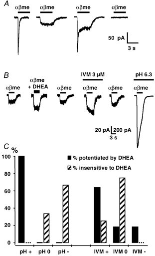
A, local application of 5 μM αβme-ATP (αβme) for 3 s induced membrane currents similar to the F-, S- and M-type currents induced by ATP (see Fig. 1A). B, illustration of the protocol used to identify the subtype of αβme-ATP-sensitive P2X receptor modulated by DHEA. We first checked for the presence (or the absence) of a potentiating effect of DHEA (50 μM) on the inward current induced by 5 μM αβme-ATP (αβme). Then in the same neurone, we determined sequentially the modulatory effect of ivermectin (IVM, 3 μM) and of an external solution at pH 6.3. The example illustrated shows a reversible potentiation of the αβme-ATP response by DHEA and IVM. Moreover, this neurone expressed αβme-ATP-sensitive P2X receptors which were strongly potentiated under low pH conditions (note the difference in calibration bar values: vertical calibration bar of 20 pA applies to the five traces on the left, whereas 200 pA applies to the last trace on the right, i.e. recorded under low pH conditions). C, correlation between sensitivity to DHEA and sensitivity to low pH (left panel) and ivermectin (IVM, right panel). Filled bars: αβme-ATP responses potentiated by DHEA; hatched bars: responses insensitive to DHEA. For each category (sensitive or insensitive to DHEA) we determined the percentage of αβme-ATP responses which were potentiated by (+), insensitive to (0) or inhibited by (−) the low pH solution or by IVM. Vh = −70 mV, n = 15.